Full Research Projects
-
[COMPLETED] Project #1 (Target Identification) - MYC Signaling in Poor Prognosis Luminal B Breast Cancer
Lead PI: Ernest Martinez, Ph.D.
Co-Lead PI: Veronica Jones, M.D.Abstract
Most luminal B breast cancers (LBBC; ER+, HER2-wt, Ki67>14%) carry a good prognosis. However, approximately 20% of LBBC are highly aggressive and resistant to current therapies. There have been many failed attempts to therapeutically target MYC. These attempts have failed, in part, due to our current inability to separate the cancer-promoting activities of MYC, mechanistically or therapeutically, from its normal essential cellular functions. In preliminary data, we show that the cancer-transforming ability of MYC is dependent on three lysine (K) residues of MYC (K149, K158, and K323) that are major substrates for acetylation by the histone acetyltransferases (HATs) p300 and GCN5. Guided by our preliminary data, here we aim to dissect the cofactors and molecular mechanisms by which these MYC acetyl-K (AcK) residues promote cell transformation and initiation and progression of LBBC. Our long-term goal is to identify new “druggable” targets to improve survival of women most affected by aggressive LBBC. Guided by our preliminary data, we hypothesize that a gene-selective MYC-AcK signaling pathway drives the aggressive tumor cell biology of therapy-resistant LBBC and involves transcription cofactors and epigenetic coregulators that “write” and/or “read” AcK marks on MYC and histones, perhaps including cofactors co-overexpressed with MYC in LBBC, such as PIN1, GCN5, p300, and/or YEATS2. Here, we will investigate the role of MYC-AcK dependent signaling in luminal mammary epithelial cell transformation and aggressive biology of LBBC. We will leverage Dr. Jones's fully annotated tissue collection of 349 women who were treated for Stage 2/3 LBBC. Aim 1 will define the impact of MYC-AcK dependent signaling in mammary epithelial cell transformation and in the aggressive biology of LBBC. Aim 2 will characterize the molecular mechanisms of MYC-AcK dependent gene regulation in transformed mammary epithelial cells and in LBBC from patients.
-
[COMPLETED] Project #2 (Drug Development) - Development of Inhibitors of the PIN1 Oncoprotein in Pancreatic Cancer
Lead PI: Maurizio Pellecchia, Ph.D.
Co-Lead: Mustafa Raoof, M.D., M.S.
Co-Investigators: Gregor Blaha, Ph.D., David Horne, Ph.D.Abstract
Pancreatic cancer is highly aggressive and has an extremely low 5-year survival rate. Pancreatic ductal adenocarcinoma (PDAC) is notoriously resistant to the majority of treatments including cytotoxic chemotherapy, targeted agents, and immuno-therapy. Treatment resistance has been linked to tumor heterogeneity, limited tissue penetration of drugs, and an immunosuppressive tumor microenvironment (TME). PIN1 is a cis-trans prolyl isomerase that controls proline-mediated phosphorylation signaling events that is overexpressed both in pancreatic cancer cells and cancer-associated fibroblasts. PIN1 overexpression is a major contributor to tumorigenesis, activating several oncoproteins, including proteins in the KRAS pathway, 17 and simultaneously inactivating several tumor suppressors. PIN1 promotes an immunosuppressive/treatment-resistant TME, by up-regulating PD-L1 (programmed cell-death receptor-1) Guided by our resources and preliminary data, we propose a collaboration between UCR and CoHCCC to optimize and develop a potent and selective PIN1 inhibitor for treatment of pancreatic cancer. Aim 1 will design, synthetize, and iteratively optimize novel, drug-like PIN1 targeting agents. Here we propose to optimize Dr. Pellecchia's initial discoveries, iteratively refining the structures of our hit compounds for potency and efficacy, with particular attention to drug-like properties of the compounds (Dr. Pellecchia, UCR). The hit-to-lead optimization phase will be fully integrated with cellular pharmacology (Dr. Pellecchia, UCR) studies, and in vitro ADME properties measurements (Dr. Horne, CoH), that will be iteratively assessed. In addition, the CoH state-of-the-art core facilities will be deployed to profile the most active agents against multiple cell lines, and to conduct high throughput cell efficacy studies in combinations. Aim 2 will study the mechanism of action and efficacy of most promising agents in cellular and animal models of pancreatic cancer. We will assess the pharmacokinetics properties of refined agents in mice and test their efficacy in animal models of pancreatic cancer.
-
Project #3 - Capacity Building Clinical Trial of Metformin Against Inflammation in Insulin-Resistant Breast Cancer Survivors
Lead PI: Kendrick Davis, Ph.D.
Co-Lead: Victoria Seewaldt, M.D.
Co-Investigator: Michelle Anne Bholat, M.D., M.P.H
Co-Investigator: Robert Rodriguez, M.D.*Previously Pilot #1
Abstract
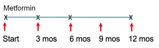
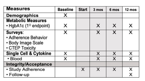
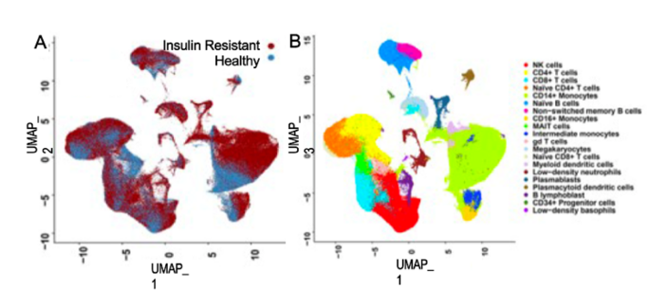
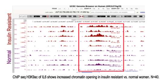
This study aims to implement a readily available, inexpensive, and safe measures to restore metabolic health and reverse accelerated aging in young adults (aged 18-35) in the rural farming Coachella Valley in Southern California. Worldwide, 34% of young adults are metabolically unhealthy, and poor metabolic health is associated with accelerated aging, decreased quality of life, and enormous economic burden for the healthcare system. Our studies show that insulin resistance leads to epigenetic damage that drives inflammation and accelerated aging. Without intervention, unhealthy young adults face a lifetime of chronic disease and early death. Effective, affordable, and scalable intervention strategies are needed to reverse metabolic disease and aging in young adults. Our proposed study will investigate the impact of metformin to reverse accelerated aging. We chose metformin because it is 1) are readily available and affordable, 2) known to restore metabolic health, and 3) shows promise to slow aging. Alone, metformin is inexpensive and safe (even in pregnancy); and it slows aging (slows telomere shortening, inflammation, DNA damage). Metformin also 1) decreases hunger by increasing sensitivity to leptin and production of GLP-1, and 2) increases delivery of glucose to muscle. Guided by our preliminary studies and supported by a concept-mapping implementation strategy, we will test in young insulin-resistant adults (aged 18-35) the hypothesis that 12-month metformin will restore metabolic health and reverse accelerated aging. We will test mechanistic markers of aging. Aim 1 will test whether metformin reverse pre-diabetes and inflammation. Aim 2 will test whether restoration of metabolic health (HgbA1c<5.7) reverse epigenetic damage and accelerated aging. Evaluate 0, 12 months. Aim 3 will test whether restoration of metabolic health (HgbA1c<5.7) improves mood and wellness using conventional measures and an Emoji evaluation tool.
-
Project #4 - Control of Protein Synthesis by eIF4A1 and mRNA m6A Modification in Acute Myeloid Leukemia
Lead PI: Seán O’Leary, Ph.D.
Co-Lead: Rui Su, Ph.D.*Previously Pilot #2
Abstract
Cancer cells require sustained high levels of protein synthesis for oncogenesis and disease progression. To achieve this, the cellular protein synthesis (“translation”) machinery is dysregulated to meet the heightened biosynthetic demands of cancer cells. Acute myeloid leukemia (AML) is a common and fatal hematopoietic malignancy. Despite improved therapeutic options, over 70% of AML patients cannot survive beyond 5 years. This underscores a critical need for more effective approaches to treat AML. Emerging evidence indicates that aberrant translation is a hallmark of leukemia and targeting mRNA translation represents a promising strategy to combat AML. Translation initiation factor 4F (eIF4F) is a heterotrimeric protein complex, including a cap-binding subunit eIF4E, an RNA-binding/scaffolding subunit eIF4G, and a DEAD-box RNA helicase eIF4A1. eIF4F recognizes the mRNA 5ʹ cap, enabling mRNA recruitment to the ribosome. This complex has been recognized as an important driver of oncogenesis; translational deregulation significantly increases its activity in cancer, including leukemia. eIF4F is thus a major cancer chemotherapeutic target. Inhibitors of all its subunits have offered significant promise as anticancer agents. N6-Methyladenosine (m6A), the most prevalent modification in mRNAs, plays a fundamental role in regulating mRNA translation, and its dysregulation might lead to oncogenesis. Building on substantial past work, we are pursuing an integrated in vivo and in vitro study that comprehensively quantifies the impact of mRNA m6A modification on eIF4A1/eIF4F function, from single-molecule level, through in vitro biochemistry, to the realm of cellular and animal physiology. Aim 1 will delineate the role of eIF4A1 in leukemogenesis and evaluate the therapeutic potential of pharmacologically targeting eIF4A1 in AML mouse models, with the goal of informing future clinical trials. Aim 2 will dissect the biochemical impact(s) of mRNA m6A, and of m6A “reader” and “writer proteins, on eIF4A1/eIF4F biochemical functions related to translation in cancer and AML. Our ultimate aim is to develop less toxic and more effective treatments for AML.
Pilot Research Projects
-
[COMPLETED] Pilot #3 - Utilizing RELM-alpha-/-(M2 Macrophage-Polarized) Mice to Develop Novel Desmoplastic Models and Targeted Therapies for Pancreatic Cancer
Lead PI: Meera G. Nair, Ph.D.
Co-Lead: Edwin R. Manuel, Ph.D.Abstract
Pancreatic ductal adenocarcinoma (PDAC) is projected to become the second-leading cause of cancer-related death by 2030. Desmoplasia, or fibrosis, is a hallmark of the PDAC tumor microenvironment (TME) and is comprised of a dense extracellular matrix (ECM) containing copious amounts of hyaluronic acid and collagens. Desmoplasia negatively affects prognosis because it: 1) acts as a biophysical barrier to therapy, 2) increases interstitial fluidic pressure leading to blood vessel compression and 3) initiates signal cascades that suppress immunity and promote invasiveness. Biochemical cues initiated by the TME ultimately contribute to proliferation, migration, invasion, angiogenesis and apoptotic resistance. Effective methods to overcome desmoplasia in PDAC, in order to improve drug delivery and efficacy, continues to be a significant unmet need. Development of novel models that physiologically recapitulate fibrosis in human PDAC would accelerate evaluation of ECM-targeting strategies. There is evidence that poor prognosis PDAC is predicted by a greater prevalence of tumor-associated macrophages (TAMs) that are primarily M2-polarized (pro-tumor) TAMs are one of the most abundant immune subsets in the PDAC stroma and promote fibrosis, tumorigenesis, immune escape, metastasis and therapeutic resistance. Strategies to eliminate or reprogram TAMs into a more M1 (anti-tumor) phenotype is currently an area of intense research. In this application, we propose complementary studies that will leverage the expertise of Drs. Meera G. Nair (UCR) and Edwin R. Manuel (COH) to develop novel tools and therapies that will aid in minimizing desmoplasia and M2 TAMs in PDAC. Aim 1 will characterize PDAC tumors implanted in M2 macrophage-driven RELM-deficient mice. Aim 2 will target M2 TAMs using a bacterial-based, shRNA plasmid delivery system.
-
Pilot #5 - Identifying Spatial Biomarkers and Therapeutic Vulnerabilities within the Native Tumor Microenvironment to Advance CAR T Cell Therapy in Glioblastoma
(previous title: Understanding CAR T Cell Efficacy in Glioblastoma Using Patient- Derived Organoids and Microenvironment-Resolved Spatial Proteoform Profiling)
Lead PI: Christine Brown, Ph.D.
Co-Lead PI: Pei Su, Ph.D.; Lisa Feldman, M.D.
Co-investigator: Xiujian Ma, M.D.Abstract
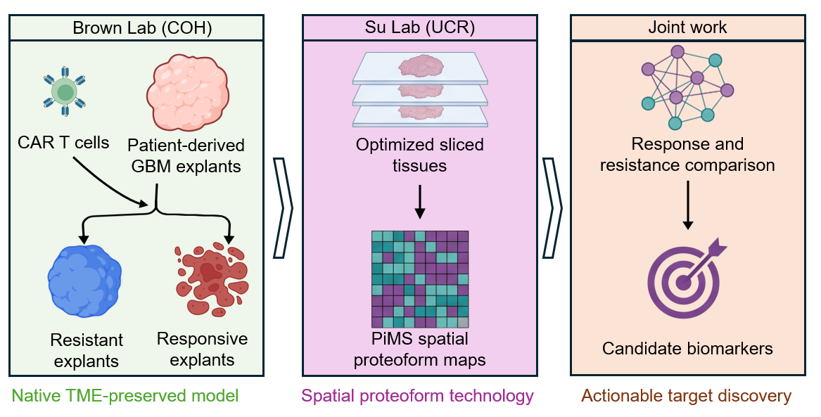
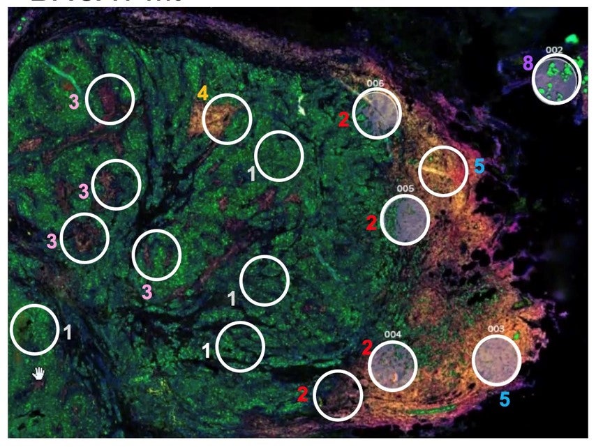
Glioblastoma (GBM) remains the most aggressive primary brain malignancy, with a median survival of approximately 15 months despite multimodal therapy. Although chimeric antigen receptor (CAR) T-cell therapy has demonstrated transformative efficacy in hematologic cancers, clinical responses in GBM remain highly heterogeneous and frequently transient. Major barriers include substantial intratumoral heterogeneity, spatially organized immunosuppressive microenvironments, limited CAR T-cell persistence, and an incomplete understanding of the molecular mechanisms underlying therapeutic resistance. Importantly, there is a lack of predictive biomarkers and actionable targets to guide patient stratification and the rational design of next- generation CAR T-cell therapies. To address this challenge, we bring together the complementary expertise of the Brown Lab at City of Hope and the Su Lab at UC Riverside. The Brown Lab has established a clinically relevant patient-derived explant (PDE) platform using a brain-mimetic PGPH hydrogel that preserves the native glioblastoma architecture, vascular components, immune populations, and tumor microenvironmental complexity while enabling the functional evaluation of clinical CAR T-cell products. Preliminary studies demonstrate heterogeneous responses across patient tumors, ranging from profound tumor killing to durable resistance, thereby providing a unique opportunity to investigate determinants of therapeutic outcome. The Su Lab has developed Proteoform Imaging Mass Spectrometry (PiMS), a next- generation spatial proteomics platform that maps intact proteoforms directly in tissues with high spatial resolution. Unlike conventional transcriptomic or antibody-based approaches, PiMS enables unbiased characterization of protein isoforms, post- translational modifications, and signaling states that directly mediate cellular function. By integrating PiMS with functional CAR T-treated PDE models, this project will establish a powerful multimodal framework linking spatial proteoform landscapes to CAR T-cell response phenotypes. Aim 1 will establish and benchmark an integrated PDE-PiMS workflow by profiling glioblastoma tissues before and after CAR T-cell treatment. We will optimize spatial proteoform analysis in PDE specimens and generate high-resolution molecular maps of responsive and non-responsive tumor regions, creating a unique atlas of CAR T- induced remodeling of the glioblastoma microenvironment. Aim 2 will leverage spatial proteomics, complementary multi-omics analyses, and multiplexed orthogonal tissue validation to identify biomarkers and therapeutic vulnerabilities associated with CAR T-cell response and resistance. We will use proteoform signatures to define cellular programs and immunosuppressive pathways enriched in resistant tumors, and nominate actionable targets that can be exploited through armored CAR T-cell engineering, combinatorial therapies, or microenvironment- directed interventions. This collaborative effort uniquely combines functional immunotherapy testing with state- of-the-art spatial proteoform discovery to move beyond biomarker identification toward mechanism-driven therapeutic development. By uncovering spatial biomarkers and targetable vulnerabilities that govern CAR T-cell efficacy in glioblastoma, this project will provide a foundation for patient stratification, rational therapeutic design, and future multi-PI NIH funding to advance next-generation cellular therapies for patients with GBM. The ultimate goal is to translate these discoveries into a first-in-human clinical trial that brings a mechanistically informed, biomarker-guided therapeutic strategy directly to GBM patients.